

Arjun Rajasekar describes how pallor detection is being used by the Raj Reddy Center for Technology and Society (RCTS) as a non-invasive method of detecting anemia.
Capitalizing on the rise of AI and the ubiquity of consumer smart devices, the Raj Reddy Center for Technology and Society (RCTS) has been exploring AI applications to improve maternal and child well-being. One of the first medical conditions chosen for exploration has been anemia, a globally prevalent issue affecting approximately 29.9% of women aged 15–49 and 39.8% of children aged 6–59 months in 2019. These rates are even higher in India, with estimates from the National Family Health Survey indicating that over 50% of women and 59% of children aged 6–59 months are anemic to varying degrees. Such widespread prevalence poses a substantial public health challenge and warrants the exploration of innovative solutions.
Anemia is characterized by a deficiency in the number of red blood cells or the hemoglobin concentration within them, resulting in a diminished capacity to transport oxygen to bodily tissues. This condition manifests through symptoms such as fatigue, weakness, dizziness, and shortness of breath. The causes of anemia are multifaceted, with iron deficiency being the most common and the easiest to remedy through supplements. Other contributing factors include vitamin B12 deficiencies, chronic diseases, genetic disorders, and bone marrow abnormalities, which may require more complex treatments.
The Need for Non-invasive Detection
Anemia is typically diagnosed through a complete blood count (CBC) test, which measures hemoglobin levels and red blood cell counts. This diagnostic procedure requires a blood sample obtained via venipuncture — an invasive and sometimes uncomfortable process that can be difficult to implement in resource-limited settings. Additionally, this method necessitates trained personnel, sterile equipment, and laboratory facilities, creating logistical barriers for vulnerable populations, particularly women and children in rural areas.
To overcome the limitations of invasive blood tests, researchers have explored various non-invasive solutions, including optical and spectroscopic devices, retinal imaging, bioelectrical impedance analysis, and pallor-based assessment. While these methods eliminate the need for drawing blood, most require dedicated hardware, making them less suitable for widespread public health screening.
Among these techniques, pallor-based assessment is the only one that does not require specialized equipment beyond what most smartphones already have — a camera. The use of pallor for anemia diagnosis has been discussed in medical literature for decades, with early studies dating back to at least the mid-20th century. The key advancement in recent years is the replacement of human observational inconsistencies with mechanized precision through computer vision (CV).
Methodology
RCTS’s framework for anemia diagnosis through pallor assessment leverages CV and AI-based image processing to analyze subtle color variations in anatomical regions traditionally examined by clinicians — such as the conjunctiva (inner eyelid), nail beds, tongue, and palm creases. Advanced object detection and segmentation techniques (YOLO and U-Net) ensure that only relevant regions (e.g., palpebral conjunctiva) are analyzed while minimizing background noise.
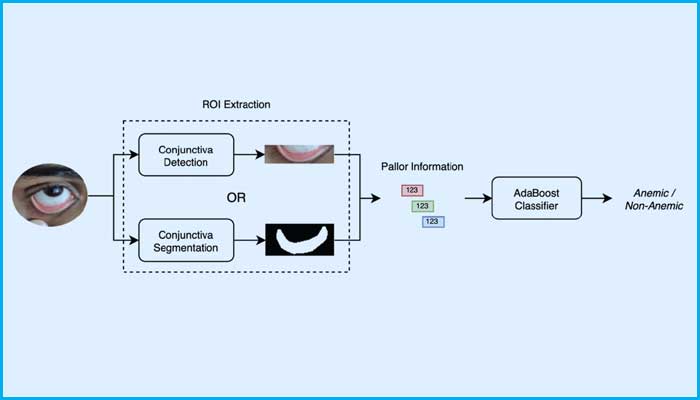
Machine learning models map pallor-related biomarkers and analyze pixel intensities correlated with hemoglobin levels to generate an anemia diagnosis. Various neural network and decision tree-based classifier architectures were explored, ultimately leading to the selection of an AdaBoost model. This ensemble model sequentially builds decision trees, with each tree focusing on correcting the errors of the previous one, ultimately combining their predictions through weighted voting.
Data Collection
The initial dataset was collected in partnership with CARE India, a non-governmental organization dedicated to promoting healthcare in rural areas. Data from approximately 2,800 patients was curated during field efforts to provide monthly health checkups for women and young mothers in Bihar. This included images of the conjunctiva, nails, palms, and tongue, along with hemoglobin level measurements when available (~1,600 patients), which served as ground truth data for anemia occurrence.
In total, ~15,000 images of the conjunctiva, nails, palms, and tongue were collected and later manually annotated for training object detection models. However, image quality varied due to field conditions and resource limitations. Since NGO operatives captured the data alongside their routine procedures, there were inconsistencies in data quality.
Results
The first phase of the study focused on a subset of the dataset with usable conjunctiva images and recorded hemoglobin values — 656 female patients (610 anemic and 46 non-anemic). The dataset was balanced using k-means SMOTE, consequent model training resulted in a binary classification model (AdaBoost) with an F1-score of ~94%. Further exploration using images of the tongue, nails, and palms is currently underway.
Uniqueness
While previous efforts to develop pallor-based anemia diagnosis systems exist, research within the Indian context has been limited to very small patient cohorts, with no publicly available datasets. RCTS aims to create an Indian-specific anemia dataset while developing more relevant models.
Limitations
The current dataset is heavily biased toward moderately anemic patients, limiting the ability to train multi-class models for anemia severity classification. Efforts are underway to collect a more demographically and diagnostically diverse dataset to validate model performance and expand its applicability. Conclusion RCTS’s exploration of non-invasive anemia detection in the Indian context through AI-driven pallor assessment presents a promising alternative to traditional blood tests, particularly for resource-limited settings. By leveraging computer vision and machine learning, this approach enhances diagnostic accessibility while minimizing the need for specialized hardware. Ongoing efforts to refine models and expand datasets will further improve the robustness and applicability of this solution for public health screening.
This article was initially published in the February ’25 edition of TechForward Dispatch

Dr. Arjun Rajasekar is an Applied Researcher at the Raj Reddy Center for Technology and Society. His areas of interest include data visualisation, predictive analytics, decision fusion, machine learning and artificial intelligence. He is passionate about developing data-driven solutions to diverse real-world problems.
Excited to see all the good work performed in RCTS. Kudos to Arjun and team.
Aravind Gondi says: