
IIITH researchers create first-ever Indian Brain Atlas demonstrating significant differences in size and structure of the average Indian brain vis-a-vis Caucasian as well as other Eastern brains. Future work in this direction is aimed at creating atlases for different age-groups to study age-related effects on brain anatomy.
When you receive the results of any pathological testing done in a lab, mere numbers don’t really convey anything. Unless they are accompanied with a reference range. To know if you’re “okay”, you typically compare your own test results against the reference range. Any deviation outside the ‘normal’ range of expected values can flag off concerns of a physiological condition or a disease.
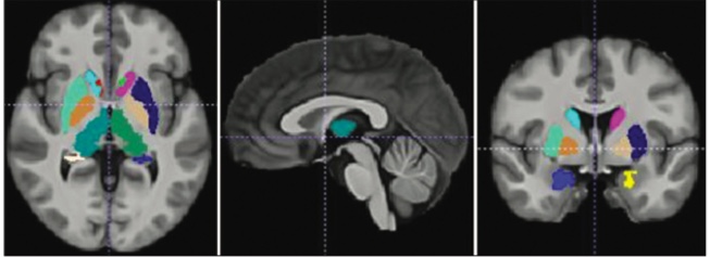
Similarly, when you undergo Magnetic Resonance Imaging (MRI) for the brain, the brain scan report needs to be compared against a ‘normal’ brain template to know if all is okay. A brain atlas or a representation of the brain made using a standard coordinate framework, allows such a comparison.
Standard Brain Atlases
The earliest known brain atlas, the Talairach and Tournoux atlas was created by manually drawing post-mortem brain sections of a 60-year-old French woman. It was in 1993 that the Montreal Neurological Institute (MNI) and the International Consortium for Brain Mapping (ICBM) created the first digital human brain atlas. More recently, MNI and ICBM have released other brain atlases that are widely used as a standard in neuroscience studies. However, these ‘standard’ brain templates created using Caucasian brains are not ideal to analyze brain differences from other ethnicities, such as the Indian population.
It was to demonstrate these inherent differences that IIITH researchers led by Prof. Jayanthi Sivaswamy from the Centre for Visual Information Technology in collaboration with the Department of Imaging Sciences and Interventional Radiology, Sree Chitra Tirunal Institute for Medical Sciences and Technology, Thiruvananthapuram, undertook the construction of the Indian human brain atlas.
Indian Brain Atlas
Speaking of the motivation behind the project, Dr. Sivaswamy says, “I’ve been working on medical imagery for a while now. And we know medical images play a big role in diagnosis. The idea of building our own Indian brain atlas came from a neuro-radiologist at the Sree Chitra Tirunal Institute. He was remarking that it is the MNI template that comes typically loaded in the MRI scanning machines, leaving us bereft of normative information.” MRI images taken are compared with the pre-loaded MNI template to arrive at a diagnosis, and likely to lead to a misdiagnosis. While even Chinese and Korean brain templates had been constructed, there was no corresponding template constructed for the Indian-specific population. The first attempt by the IIITH team at creating an Indian-specific brain atlas involved 50 subjects, evenly balanced out across genders. MRI scans of these subjects’ brains were taken at three different hospitals across three different scanners to rule out variations found in scanning machines. Emboldened by the results of the pilot study, the team went on to recruit 100 willing participants in the eventual construction of the Indian Brain Atlas, referred to as IBA 100.
From birth, the brain grows at an alarming rate. But according to most experts, it is around age 20-30, that the brain is said to be fully developed, or ‘mature’. Hence the scans collected were from an equal number of healthy male and female subjects who fell in the age group of 21-30 years, considered as the baseline.
Results
The constructed altas was validated against the other atlases available for various populations. It was found that the Indian brain on average is smaller in height, width, and volume as compared to the western (MNI) as well as the eastern population (Chinese and Korean). These differences are found even at the structure level, such as in the volume of the hippocampus and so on. But overall, the IBA 100 is more comparable to the Chinese and Korean atlases than the distant Caucasian one (MNI).
Future Work
Dr. Sivaswamy admits that it is desirable to build a larger atlas with a greater heterogeneous mix of subjects to account for diversity, even in terms of educational qualifications. But currently the team’s focus is to understand the aging process itself. “There are many changes that take place in a brain due to advancing age, with the most typical one being atrophy. Atrophy means shrinking of structures. In the case of dementia or Alzheimer’s, they are associated with atrophy of the hippocampus,” explains Dr. Sivaswamy. For this, her PhD student, Alphin Thottupattu is collecting MRI scans to create brain atlases for different age groups, like 20-30, 30-40, 40-50 and 50-60. This will be beneficial to track the brain and see how it ages over time. While others in the scientific community are currently undertaking a longitudinal study of the ageing effects on the brain, IIITH’s approach is likely to showcase results faster and demonstrate variability too. “We may primarily be a young country but with increasing mortality, the number of aged persons is increasing too. And so are incidences of Alzheimers and dementia. It is important to understand structurally what is normal too, so we can catch such conditions early on. Such research has a lot of social impact,” says Dr. Sivaswamy.

Sarita Chebbi is a compulsive early riser. Devourer of all news. Kettlebell enthusiast. Nit-picker of the written word especially when it’s not her own.
Please provide more details like which MRI machine was used, how many Tesla, with or w/o anesthesia, contrast, etc. Can you cite the publication?
Suresh says: